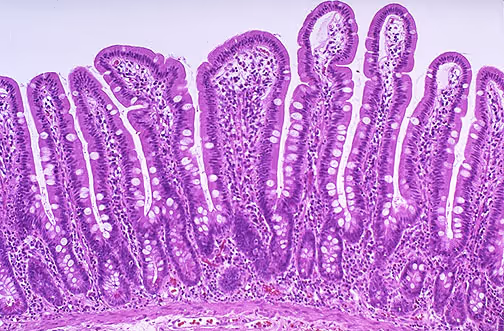
Spatial multi-omics has the potential to reveal intricate patterns and beautiful panoramas of gene and protein expression, which can improve our understanding of biology, enable better biomarker discovery, and support identification of new mechanisms of disease and drug action.
Considering the advances the field has seen and the release of commercial instrumentation in recent years, it is no surprise that at least one quarter of the abstracts at this year’s Advances in Genome Biology and Technology (AGBT) meeting were devoted to spatial biology. The industry is keen to serve this promising market.
Companies are mostly pursuing two main technical approaches, FISH – short for in situ fluorescent hybridisation – and sequencing, to identify tissue transcripts.
With the current cost of sequencing, FISH offers a more affordable route to profiling the expression of panels of known genes of interest with exquisite, subcellular resolution. The focus on known biomarkers also offers an obvious path for FISH to enter the clinic.
Spatial sequencing, in contrast, makes it possible to study gene expression without prior knowledge of the genes of interest, making it ideal for true de novo discovery applications. But, for now, this approach is still too expensive, and it is too challenging to practically extend “spatial ‘omics” via sequencing to single-cell resolution.
With so many players in spatial biology, both FISH-based technologies and spatial sequencing companies will need to cement their position in this market.
In this blog, we discuss the scope for innovation in the spatial biology market.
In situ fluorescent hybridisation (FISH)
In FISH, fluorescently labelled nucleic acid probes are combined with sophisticated imaging technologies to detect and quantify nucleic acids in cellular environments.
In recent years, spatial biology companies have further combined FISH with advances in microfluidics to automate the sequential detection of handfuls of targets. This approach can be used to build up an image consisting of up to 1000 probes to visualise spatial patterns of gene expression. Gene expression matrices can also be overlaid with standard tissue section images to correlate gene expression with phenotype.
FISH-based approaches also make it relatively straightforward to detect proteins of interest by combining nucleic acid-based probes and antibodies with a molecular identification barcode. Probe panels can be designed for multi-omics investigations, for example of an entire pathway. FISH could also be used to visualise cellular functions such as RNA splicing or potentially even genomic rearrangements where they are suspected to be involved.
Moving from the biology to the clinic, FISH also shows good performance with formalin-fixed paraffin-embedded (FFPE) tissue samples – the majority of clinical samples. This is because probe-based approaches can be designed to target any part of a transcript, avoiding the 3’ end, to which RNA degradation is biased. FISH technologies can effectively probe very degraded mRNA that would often be dismissed as unusable by the sequencing community (RNA integrity number <3 are frequently observed for FFPE).
However, with current technologies each probe detection cycle takes significant time for reagent delivery, probe hybridisation, washing and image acquisition. Improved biology and fluidic innovation could increase the number of targets acquired per cycle and reduce cycle lengths. However, this may quickly reach a ceiling where instrumentation cost and fluidic complexity spiral out of control.
Ultimately, the strength – but also the obvious drawback – of FISH is its targeted nature. It works with established targets, and this may provide a more obvious route into the clinic. But ultimately, this limits the utility of FISH for true de novo discovery applications.
Spatial sequencing
Sequencing has been applied more recently as an alternative approach for visualising gene expression across a tissue section, also known as “spatial transcriptomics”.
RNA transcripts from the tissue section are captured on a microscope slide seeded with a “lawn” of conjugated capture probes, similar to a microarray. The coordinates of captured molecules each possess a unique spatial barcode, which is integrated into the library molecules during first strand synthesis, enabling the user to later bioinformatically reassign reads to the spatial coordinates from which they came.
In sequencing-based technologies, transcripts are often captured by probing their poly-A tail, which is present in all intact mRNA transcripts. Sequencing can therefore capture any transcript present without any prior identification. While there are at least 23,000 genes in the human genome, and many more transcript species, FISH can capture only on the order of 100s-1,000s at a time. This makes sequencing-based methods ideal for discovery applications.
The full power of sequencing could be unleashed in drug discovery and fundamental research as it allows users to sequence deep enough to identify rare species. In addition, sequencing-based approaches are quantitative, supporting statistical analysis and comparison across studies to give meaningful biological patterns.
And whereas FISH can only detect the main four DNA/RNA bases, sequencing protocols can also quantify epigenetic marks and splice variants across the genome. And at the moment only a small number of companies are active in this market – making it an attractive space for innovation.

Scope for innovation
Sequencing is still too expensive to match the single-cell resolution offered by FISH. But seeing the promise of declining sequencing costs and the emergence of new companies and products in the market, recent IP agreements [1], and Ultima Genomics’ (and others) push for the $100 genome, this market may present opportunities for the industry sooner than one might expect.
Companies with an eye on this market can use the interim to solve major technology challenges. At the time of writing, barcode printing methods on the market only offer ~10-cell resolution. However, replacing droplet-based printing with other approaches that open up micron or sub-micron scale resolution is eminently possible.
At TTP, we are exploring alternative approaches to enable single-cell spatial multi-omics, without increasing consumable costs or cumbersome manufacturing processes. Innovation in biochemistry (e.g. barcoding methodologies), fluidics, optics, and the application of computational tools in concert are significant facilitators in the race to improve resolution without escalating costs for the user.
Currently, sequencing-based methods usually rely on intact mRNA molecules being present, because they capture transcripts by targeting the poly-A tail. But in FFPE samples, for example, this is often damaged due to enzymatic and mechanical degradation during storage. Inventive methods are needed to increase the sensitivity in such samples without biasing sequencing-based approaches towards the most stable or abundant transcripts, or resorting to targeted panel-based sequencing.
In the meantime, there is scope to cement FISH-based technologies in the market with improvements, particularly with the clinic in mind.
Increased throughput is essential for diagnostic and drug discovery applications of FISH-based technologies. The fastest available implementations analyse a modest batch of samples in a day(s), making this the current gold standard.
Improvements can be achieved by rethinking reagent delivery and image acquisition technologies – for example by developing optimised flow cell geometries and innovative fluid handling technologies that minimise reagent use and maximise delivery speed.
Detection is currently limited by challenges associated with panel design for simultaneous target detection and the cost of highly multiplexed optical systems to interrogate several different fluorophores at one time. Improved reagent design allowing highly multiplexed panels to be used alongside simplified optical design would be a significant differentiator in this field, enabling higher plex panels without escalating instrument costs.
Cost and user uptake
The true differentiators, however, will be cost, speed and flexibility, which together will drive user uptake.
To dominate the market, companies need to achieve instrument sales in the low thousands range, which, in turn, will drive revenue from consumables. Achieving this could hinge on accessible features, meaningful results, cost, throughput and, for the clinic, compliance with regulatory standards.
While features such as 1000-plex RNA and 100-plex protein panels are now common for FISH-based technologies, additional multi-omics capabilities such as the detection of DNA and RNA epigenetic marks, splice variance and accurate transcript quantification could increase the value and adoption of any technology.
For FISH technologies and targeted sequencing-based technologies, available panels are still limited to human and mouse models, whereas discovery biology is often done in other model organisms – leaving a potential market unaddressed.
As has been the experience in the single-cell sequencing field, accessible analytical tools can be essential in building customer loyalty. Efficient handling of the huge datasets (>300 GB) can significantly alleviate the burden placed on the users and provide the needed novel insights in a short amount of time.
As we have seen in the sequencing market, instrument cost does play a big role in customer uptake. By cost engineering in instrument design processes, coupled with innovation in reagent design, burden on costly instrument features such as high-resolution multi fluorescent channel optics can be reduced. With the promise of lower sequencing costs, sample prep and reagent costs will also likely come into the spotlight.
At the end of the day, if a company wants to enjoy long-term success in drug discovery and diagnostic applications or get favoured by big academic facilities, strategic market-oriented thinking will be essential.
While it is likely that both FISH-based spatial technologies and the application of spatial sequencing will continue to play a role, hitting the right combination of biological insight, instrument usability, and cost will be key to building a technology to lead the spatial biology market.
Find out how we help clients in Diagnostics and Life Science.
You can also read our previous related blog post: Next-generation sample preparation: Will better nucleic acid extraction unleash biotechnology, life sciences and diagnostics?


